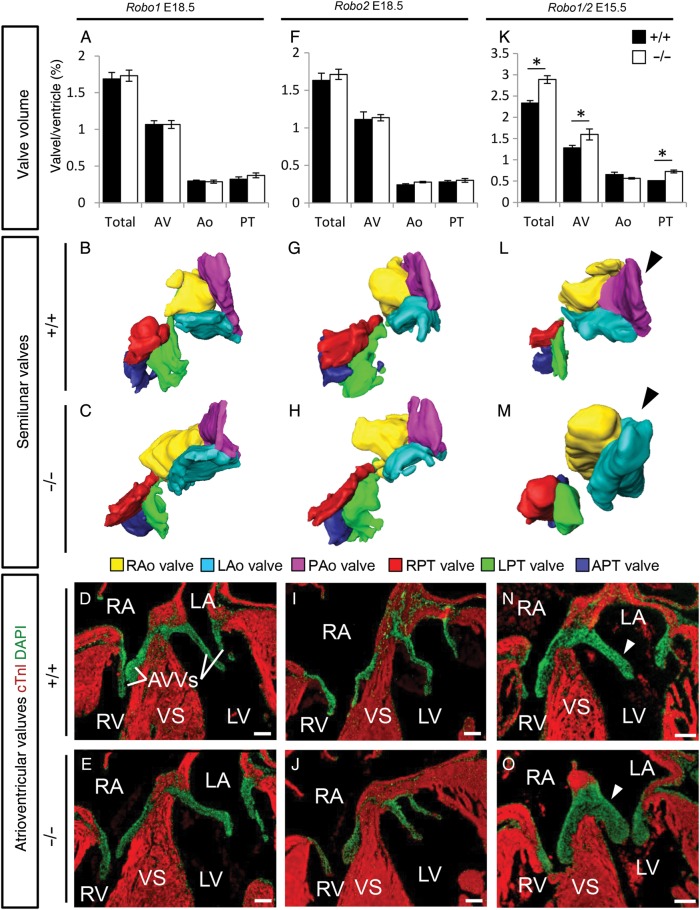
Figure 4.
A spectrum of valve malformations in Robo mutants. (A–O) Analysis of the cardiac valves at the indicated developmental stages in Robo1+/+ (A, B, and D) Robo1−/− (A, C, and E) Robo2+/+ (F, G, and I) Robo2−/− (F, H, and J) Robo1+/+;Robo2+/+ (K, L, and N), and Robo1−/−;Robo2−/− (K, M, and O) embryos. (A, F, and K) Measurements of the total valve (Total), atrioventricular valve (AV), aortic valve (Ao), and pulmonary trunk valve (PT) volume corrected for ventricular volume. (A) Total, n = 5, P = 0.47; AV, P = 0.60; Ao, P = 0.75; PT, P = 0.35. (F) Total, n = 5, P = 0.52; AV, P = 0.47; Ao, P = 0.08; PT, P = 0.92. (K) Total, WT n = 5, KO n = 3, P = 0.025; AV, P = 0.025; Ao, P = 0.30; PT, P = 0.025; Mann–Whitney U test. Note the overall increased valve volume in Robo1−/−;Robo2−/−. (B, C, G, H, L, and M) Example three-dimensional reconstructions as used for the volume measurements of the semilunar valves, seen from the ventricular side. Black arrowhead, note the absence of the posterior aortic valve in the Robo1−/−;Robo2−/− (M) embryo. (D, E, I, J, N, and O) Examples from immunohistochemistry sections (cTnI and DAPI) used for the measurements, showing the atrioventricular valves. White arrowheads, Robo1−/−;Robo2−/− (O) embryos show thickened valves. R/L/PAo, right/left/posterior aortic valves; R/L/APT, right/left/anterior pulmonary trunk valve; AVVs, atrioventricular valves. *P < 0.05. For other abbreviations, see the legend of Figures 1 and 2. Scale bars depict 100 µm.

