
Figure 5.
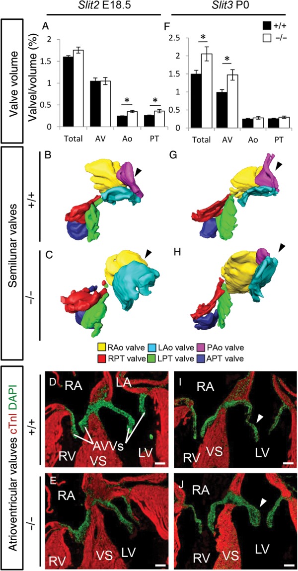
A spectrum of valve malformations in Slit mutants. (A–J) Analysis of the cardiac valves at the indicated developmental stages in Slit2+/+ (A, B, and D), Slit2−/− (A, C, and E), Slit3+/+ (F, G, and I), Slit3−/− (F, H, and J) embryos. (A and F) Measurements of the total valve (Total), atrioventricular valve (AV), aortic valve (Ao), and pulmonary trunk valve (PT) volume corrected for ventricular volume. (A) Total, n = 5, P = 0.08; AV, P = 0.92; Ao, P = 0.016; PT, P = 0.025. (F) Total, WT n = 5, KO n = 3, P = 0.025; AV, P = 0.025; Ao, P = 0.46; PT, P = 0.10; Mann–Whitney U test. Note that while there is overall increased valve volume in Robo1−/−;Robo2−/− mutants, Slit2−/− mutants only show increased semilunar (A) and Slit3−/− only increased total and atrioventricular valve volume (F). (B, C, G, and H) Three-dimensional reconstructions of the semilunar valves, seen from the ventricular side. Black arrowhead, note the absence of the posterior aortic valve in the Slit2−/− (C) while this valve is hypoplastic in the Slit3−/− (H) embryos. (D, E, I, and J) Immunohistochemistry sections (cTnI and DAPI) showing the atrioventricular valves. White arrowheads, Slit3−/− (J) embryos show thickened valves. R/L/PAo, right/left/posterior aortic valves; R/L/APT, right/left/anterior pulmonary trunk valve; AVVs, atrioventricular valves. *P < 0.05. For other abbreviations, see the legend of Figure 1. Scale bars depict 100 µm.