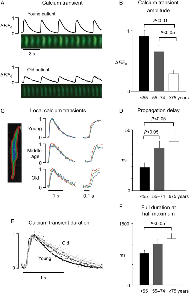
Figure 1.
Effects of ageing on the intracellular calcium transient. (A) Calcium transient traces and the corresponding sequence of 68 consecutive time-averaged calcium images, recorded in myocytes from a young and an old patient. (B) Average calcium transient amplitude in myocytes from young (8 cells; n = 7) middle age (10 cells; n = 7), and old (6 cells; n = 5) patients. (C) Calcium transients measured in three concentric rings (shown on the left). Transients were normalized to their peak values in a young, a middle aged, and an old patient. The upstroke of the calcium transient is amplified on the right. Notice the delay between the calcium transient near the sarcolemma (SL; red) and the cell centre (CC; blue) in the old patient. (D) Average time-delay between the calcium transient in the SL and CC for the three patient groups. (E) Superimposed calcium transients normalized to their peak amplitude. (F) Average duration of the calcium transient at half maximal amplitude. P-values for significant differences are indicated above the corresponding bars.

