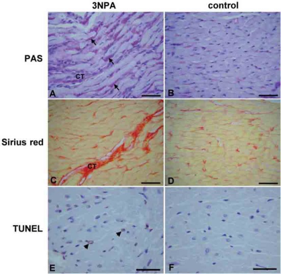
FIGURE 1.

Histopathological changes in hearts of rats treated with 3NPA (A, C, E) as compared with control rats treated with normal saline (B, D, F). In 3NPA-treated animals PAS staining (A, B) showed increased amount of glycogen granules in the cytoplasm of cardiomyocytes (arrows), Sirius red staining (C, D) showed myocardial fibers interrupted with increased amount of connective tissue (CT), while TUNEL staining (E, F) showed sparse TUNEL positive cells (arrow-heads). bar (A, B, C, D) = 50 μm; bar (E, F) = 30 μm.
