Figure 3.
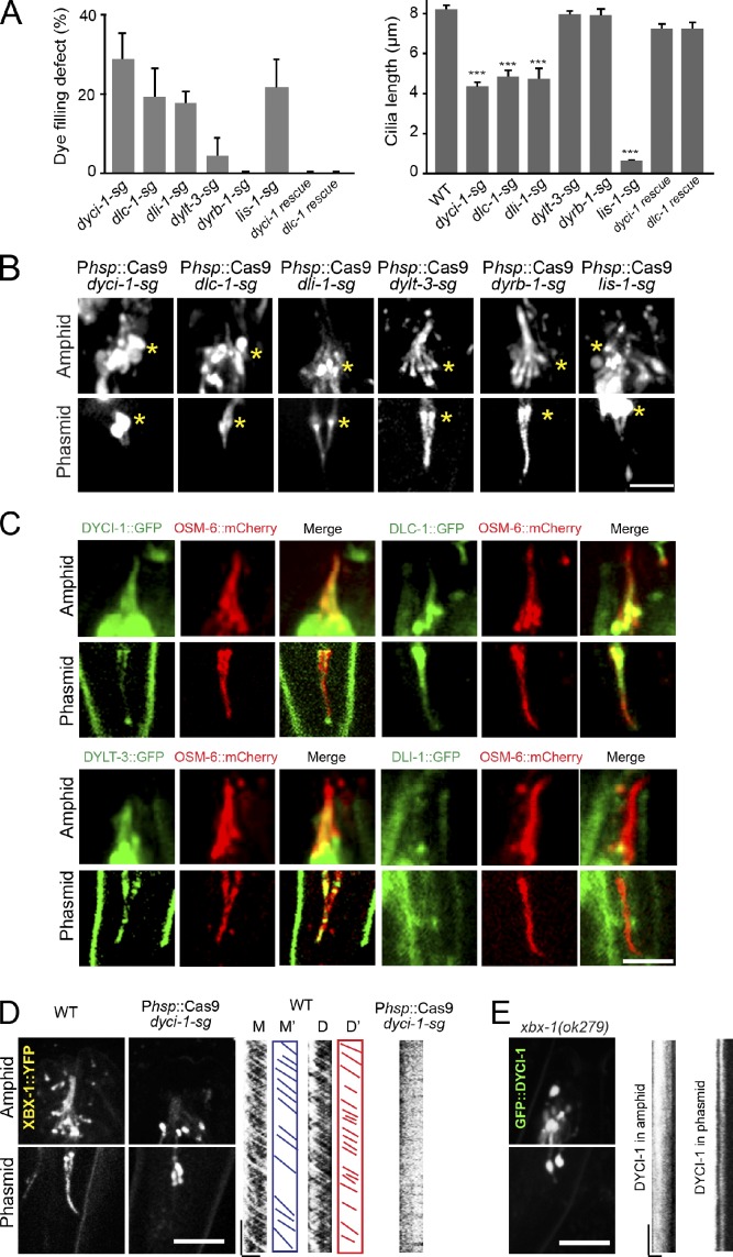
Ciliary defects in conditional mutants of embryonically essential dynein components. (A) Defects of dye-filling assays (left) and the cilium length (right). Mean ± SE (error bars); n = 34–381 from three generations. ***, P < 0.001. (B) Amphid and phasmid cilia visualized by OSM-6::GFP. Asterisks indicate transition zones. Bar, 5 µm. (C) Ciliary localization of DYCI-1, DLC-1, DYLT-3, and DLI-1 (GFP) and OSM-6::mCherry (red). (D and E) Localization (left) and motility (kymographs, right) of XBX-1::YFP in WT and dyci-1 conditional mutants (D) or GFP::DYCI-1 in xbx-1 mutants (E). Micrograph bar, 5 µm; kymograph horizontal bar, 2 µm; vertical bar, 5 s.

