Figure 2.
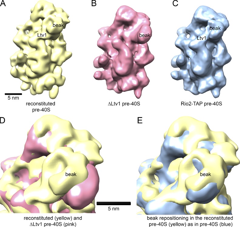
ΔLtv1 pre-40S ribosomes reconstituted with Yar1–Rps3 and Ltv1/Enp1 are structurally identical to native pre-40S ribosomes. (A) The solvent face of the reconstituted pre-40S ribosome shows the features expected from the presence of Enp1/Ltv1 and Rps3 near the beak. (B) Identical view of the ΔLtv1 pre-40S ribosomes used as a starting material in reconstitutions (EMD1924; Strunk et al., 2011). (C) Natively purified pre-40S ribosome (EMD1927; Strunk et al., 2011). (D) A superimposition of reconstituted (yellow) and ΔLtv1 pre-40S (pink) shows the repositioning of the beak upon addition of Enp1–Ltv1–Rps3, which suggests a structural recapitulation of the biochemically characterized pre-40S state. (E) Superimposition of reconstituted (yellow) and natively purified (blue) pre-40S ribosomes shows that reconstituted pre-40S ribosomes are structurally identical to purified native pre-40S ribosomes.

