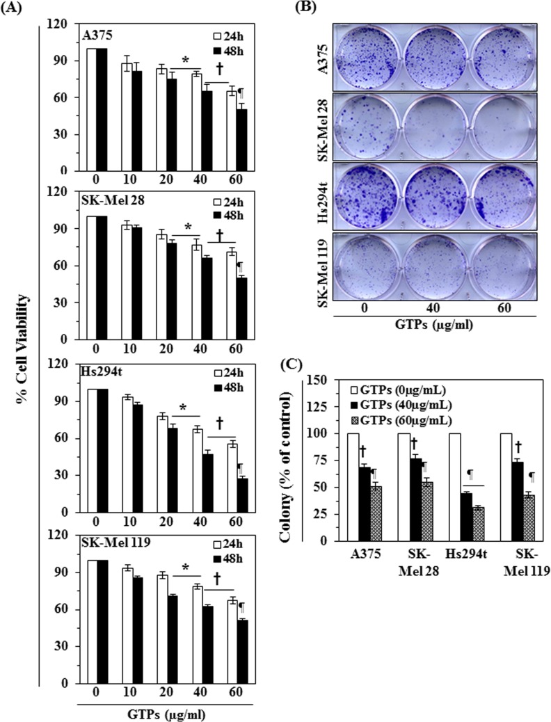
Figure 1. Cytotoxic effect of GTPs on melanoma cells.
(A) Treatment of human melanoma cells (A375, SK-Mel28, Hs294t, SK-Mel119) with various concentrations of GTPs (0, 10, 20, 40 and 60 μg/ml) inhibits the proliferation or cell viability in a dose- and time-dependent manner. Cell viability was determined using MTT assay as described in the Materials and Methods section, and data are expressed in terms of percent of control group (non-GTPs treated) as the mean ± SD of six replicates. Significant difference versus non-GTPs-treated controls, *P <0.05; †P<0.01; ¶P<0.001. (B) Treatment of melanoma cells with GTPs for 2 weeks inhibits the colony formation ability of cells. Cancer cell colonies are shown in blue-purple. (C) Number of colonies in each treatment group was detected and counted under Olympus microscope and data on colony formation are summarized in terms of percent of control. Significant difference versus control, †P<0.01, ¶P<0.001.

