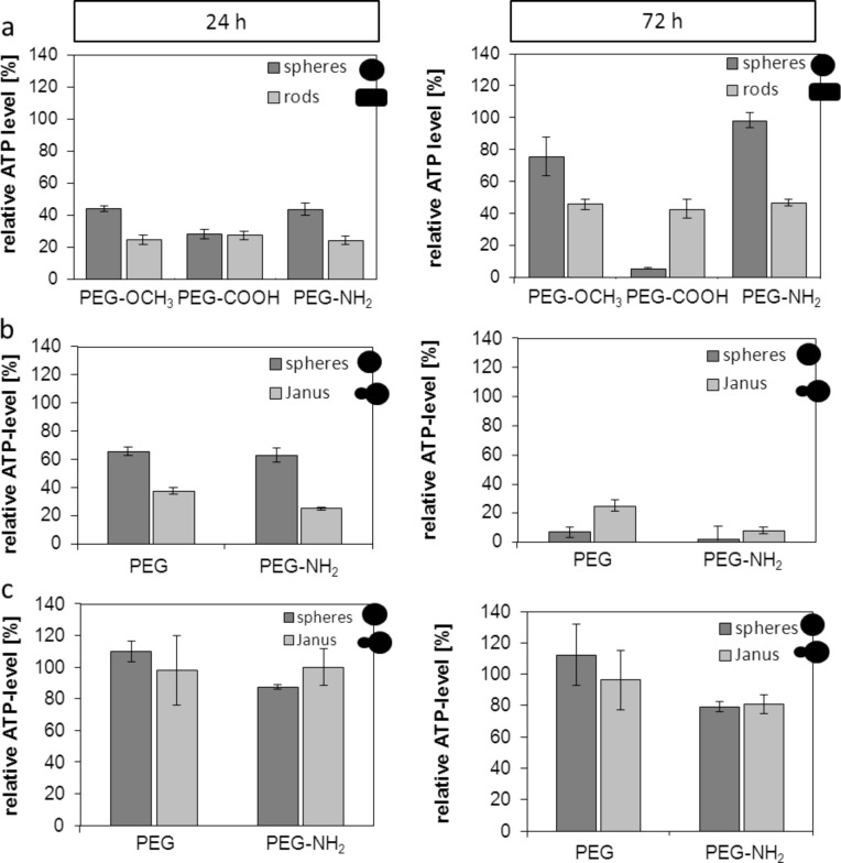
Figure 1.
Impact of different shaped and functionalized nanoparticles on the cellular ATP-level of different endothelial cells after 24 h and 72 h of incubation. Relative cellular ATP-levels were detected by ATPLite assay. (a) SVEC4-10 were treated with 30 µg/mL of gold nanoparticles functionalized with OCH3, COOH or NH2. (b) HMEC-1 cells were treated with 20 µg/mL of MnO and Au@MnO nanoparticles. (c) HMEC-1 cells were treated with 20 µg/mL of Fe3O4 and Au@Fe3O4 nanoparticles. Data were normalized to control values (no particle exposure), which were set to 100% ATP level.

