
Fig. 3.
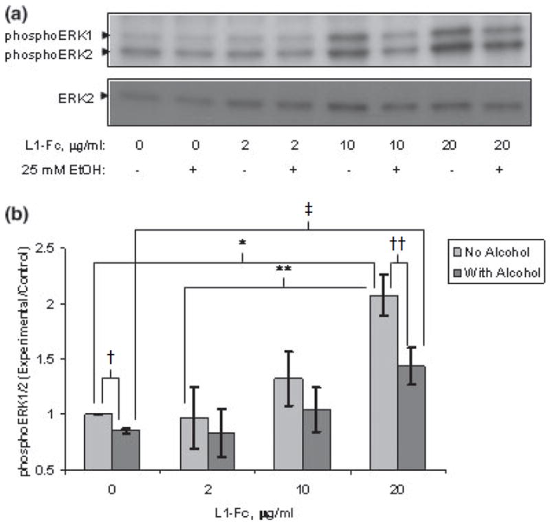
(a) Ethanol inhibits L1-Fc activation of ERK1/2. Cerebellar granule cells grown in serum-free defined media were treated with or without 25 mm ethanol for 1 h prior to addition of L1-Fc at increasing concentrations. ERK1/2 activation (phosphoERK1/2) was assayed by western blot analysis after addition of L1-Fc or vehicle alone. A representative blot of four experiments is shown. (b) Ethanol significantly inhibits L1-Fc activation of ERK1/2. Densitometric quantification of ERK1/2 phosphorylation corrected for total ERK2 shown in (a) is plotted as relative densitometric units relative to the vehicle alone control. The values of four separate experiments are shown. The bar indicates the mean of the values ± SE. Addition of 20 μg/mL of L1-Fc significantly increased phosphoERK1/2 above both addition of vehicle alone (*p < 0.005, paired t-test) and 2 μg/mL L1-Fc (**p < 0.002, paired t-test). Ethanol pretreatment of 25 mm for 1 h prior to addition of L1-Fc significantly reduced phosphoERK1/2 both in the vehicle alone (†p < 0.001, paired t-test) and 20 μg/mL L1-Fc-added cells (††p < 0.0004, paired t-test). PhosphoERK1/2 was significantly greater in the 20 μg/mL L1-Fc + ethanol cells compared with the vehicle alone + ethanol cells (‡p < 0.024, paired t-test).