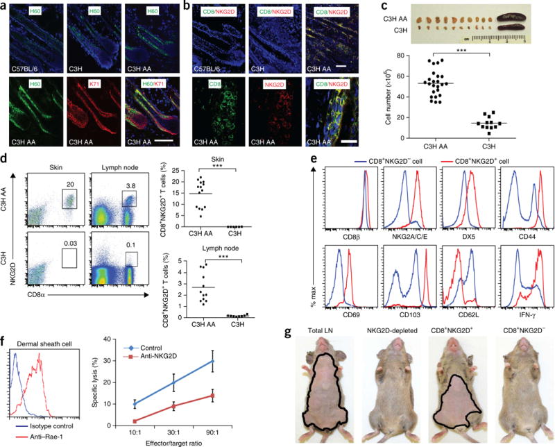
Figure 1.

CD8+NKG2D+ cytotoxic T lymphocytes accumulate in the skin and are necessary and sufficient to induce disease in AA mice. (a) Immunofluorescence staining of NKG2D ligand (H60) in the hair follicle inner root sheath (marked by K71). Scale bar, 100 μm. (b) CD8+NKG2D+ cells in hair follicles of C57BL/6, healthy C3H/HeJ and C3H/HeJ AA mice. Top scale bar, 100 μm; bottom scale bar, 50 μm. (c) Cutaneous lymphadenopathy and hypercellularity in C3H/HeJ AA mice. (d) Frequency (number shown above boxed area) of CD8+NKG2D+ T cells in the skin and skin-draining lymph nodes in alopecic mice versus ungrafted mice. (e) Immunophenotype of CD8+NKG2D+ T cells in cutaneous lymph nodes of C3H/HeJ alopecic mice. (f) Left, Rae-1t–expressing dermal sheath cells grown from C3H/HeJ hair follicles. Right, dose-dependent specific cell lysis induced by CD8+NKG2D+ T cells isolated from AA mice cutaneous lymph nodes in the presence of blocking anti-NKG2D antibody or isotype control. Effector to target ratio given as indicated. Data are expressed as means ± s.d. (g) Hair loss in C3H/HeJ mice injected subcutaneously with total lymph node (LN) cells, CD8+NKG2D+ T cells alone, CD8+NKG2D− T cells or lymph node cells depleted of NKG2D+ (5 mice per group). Mice are representative of two experiments. ***P < 0.001 (Fisher’s exact test). For c,d,f, n and number of repeats are detailed in the Supplementary Methods.
