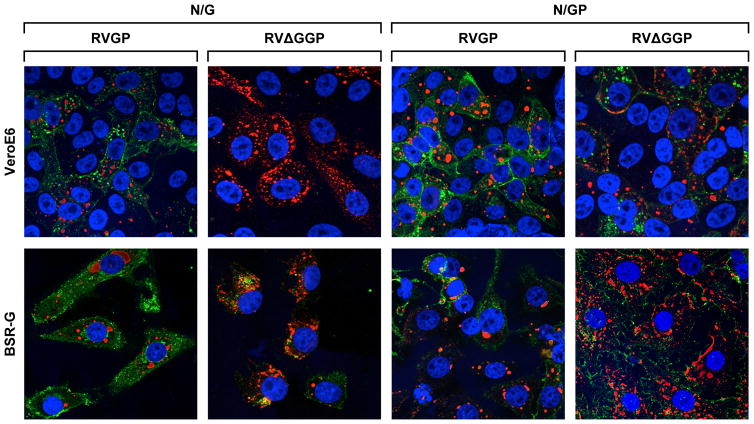
Figure 3.
Confocal microscopy at 126X magnification using fluorescently labeled antibodies shows that RVG is not synthesized in RVΔGGP-infected VeroE6 cells. BSR-G and VeroE6 cells were infected with RVGP or RVΔGGP at an MOI of 0.01, and then fix and stained at 48hpi according to staining method in Table 1. Antibody set N/G is for RABV N and RABV G detection; N is red and G is green. Antibody set N/GP is for RABV N and EBOV GP detection; N is red and GP is green.

