FIGURE 5.

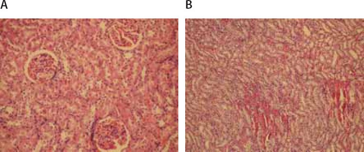
(A) Glomeruli and tubular cells showed normal appearance (Hematoxylin–eosin × 200). (B) Stasis, congestion and hemorrhage in some sections of medulla (Hematoxylin–eosin × 100).

(A) Glomeruli and tubular cells showed normal appearance (Hematoxylin–eosin × 200). (B) Stasis, congestion and hemorrhage in some sections of medulla (Hematoxylin–eosin × 100).