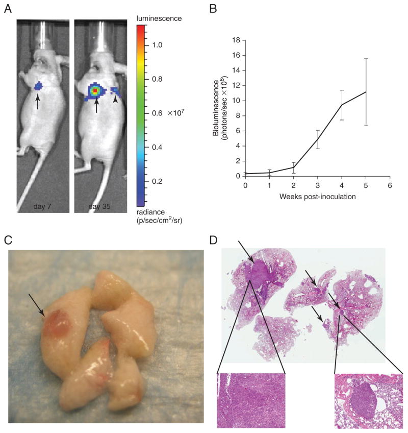
Figure 14.27.2.
Monitoring of tumor growth in orthotopic murine model of NSCLC by in vivo bioluminescence imaging. (A) Lateral view of bioluminescent images of mouse bearing lung orthotopic tumor of H1299. The H1299/Luc cells (1 × 10⁶) were inoculated into the left lung of nude mice. (Left) Image of a primary H1299 tumor in the left lung at 7 days after implantation. (Right) Both primary lung tumor growth and presence of a metastatic lesion in the contralateral lung are evident 35 days after implantation. (B) Quantitative analysis of luciferase bioluminescence images taken at the indicated time points. Data are mean total bioluminescence flux (photons/sec) ± SEM. (C) Macroscopic view of H1299/Luc tumor (arrow) on lung parenchyma. (D) Hematoxylin and eosin staining of harvested mouse lung demonstrate primary tumor in left lobe (thick arrow) and multiple metastases in contralateral lobes (thin arrows) (1× magnification). Insets: Higher (5×) magnification of primary tumor and metastasis.

