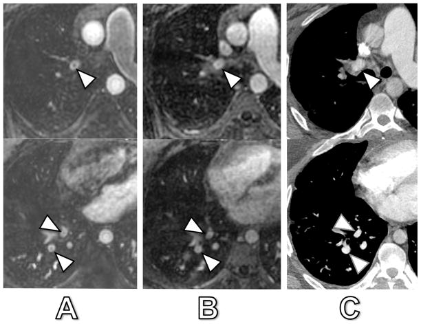
Fig. 3. 31-year old man with signal drops in lobar and segmental pulmonary arteries.
Arterial-phase MRA (A), delayed-phase MRA (B) and corresponding CT (C). The patient had no acute embolism by CT. The central signal dropout in the right upper lobe lobar artery (arrowhead) corresponded to artifact with a 51% and 23% signal drop at arterial-phase and delayed-phase MRA, respectively. The central signal dropout in the right lower lobe segmental arteries (arrowheads) corresponded also to artifact with a 41% and 18% signal drop at arterial-phase and delayed-phase MRA, respectively.

