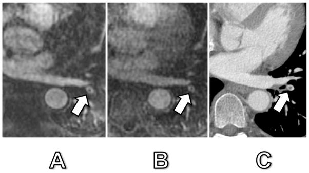
Fig. 4. 42-year old woman with confirmed PE.

Arterial-phase MRA (A), delayed-phase MRA (B) and corresponding CT (C). The central signal drop in a left lower lobe segmental pulmonary artery (arrow) corresponded to a true pulmonary embolus as confirmed by CT. Signal dropout was 57% and 52% at arterial-phase and delayed-phase MRA, respectively. Of note, this embolus was the only one that was detected in this patient and could easily have been mistaken for a truncation artifact due to its central location within the vessel and relatively small signal intensity drop.
