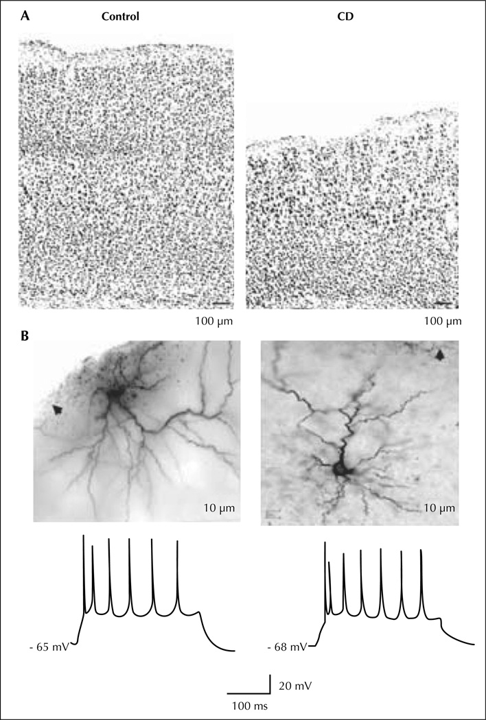
Figure 5.
In utero irradiated rat model of cortical dysplasia. Pyramidal cells from the dysplastic cortex (CD) are spatially disorganized and dysplay a regular spiking pattern to suprathreshold current pulses. A) Low power photomicrographs of coronal sections of control (left) and dysplastic (right) cortex with cresyl violet staining. B) Photomicrographs (upper) show two biocytin-stained pyramidal cells from dysplastic cortex. Arrows point to pia. Traces (lower) show spike patterns of these two cells to depolarizing current injection (300 ms, 300 pA).

