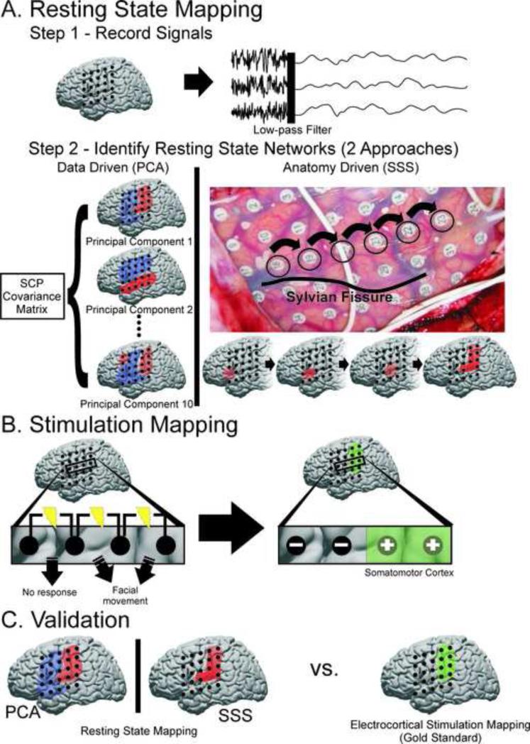
Fig. 1.
Schematic for identifying SCP correlation networks and testing their performance against the gold standard for delineating SM cortex. A) Step 1, resting-state electrical activity from both awake and anesthetized patients was recorded from cortical electrodes, re-referenced, and filtered for the SCP (<0.5Hz). Step 2, two approaches were taken to identifying candidate SCP correlation networks hypothesized to reflect SM cortex. The first was a data driven approach in which principal component analysis was applied to the covariance matrix of the SCP signals from each electrode. The top 10 principal components (illustrated in blue and red) were considered possible candidates for SM cortex. The second was an anatomy based approach in which seeds were selected superior to the Sylvian fissure in an anterior-posterior direction to maximize likelihood of intersecting SM cortex and identifying the corresponding SCP correlation network. B) ECS mapping was used as the “gold standard” to define SM cortex. Electrodes were labeled as ECS negative for SM cortex if they were involved in any bipolar stimulation which did not elicit a motor or sensory response. Electrodes were labeled as ECS positive for SM cortex if they were only associated with a motor or sensory response when involved in a bipolar stimulation. C) SM cortex defined by ECS mapping was used to assess the sensitivity and specificity of the new techniques based on resting state SCP correlation networks.

