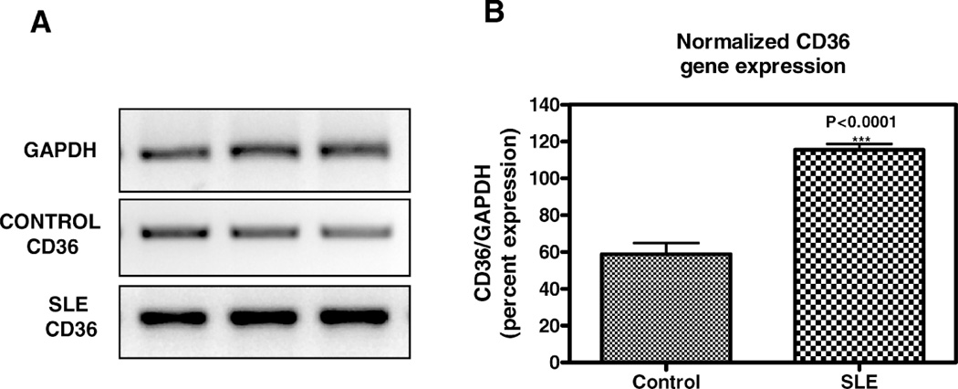
Figure 2.
Effect of SLE plasma versus normal human plasma on CD36 mRNA in THP-1 monocytes. THP-1 monocytes were incubated in 50% volume/volume SLE patient or normal control plasma for 3 hours as indicated. Following incubation, total RNA isolated from cells exposed to each condition was reverse transcribed and amplified by PCR with GAPDH message as an internal standard. A) Representative experiment from a total of three SLE patients and three controls studied. Photograph of ethidium bromide-stained PCR-amplified bands corresponding to message for CD36 and GAPDH as indicated. B) Gene expression levels were graphed as relative mRNA expression. The data represent the mean and SEM of three independent experiments.

