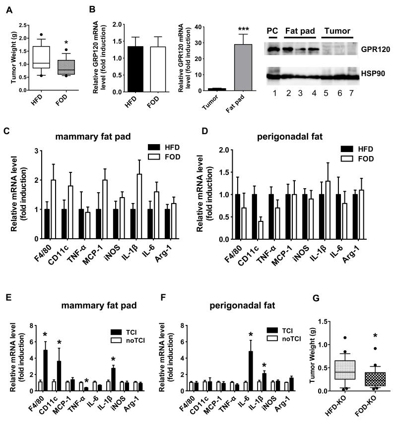
Figure 5. FOD significantly reduces mammary tumor growth in both WT and GPR120 KO mice and tumor cell injection modulates inflammation in the mammary fat pad and perigonadal fat.
A: OVX WT mice were fed a HFD for four weeks and then either switched to a FOD or maintained on the HFD. Py230 cells were injected into the mammary fat pads at 14 weeks after starting the HFD and tumor burden was measured at 8 weeks after tumor cell injection (22 total weeks of diet protocol, n=16 tumors per group). *p < 0.05 vs. HFD. B: The mRNA levels for GPR120 in mammary tumors and the mammary fat pad of HFD- and FOD-fed mice were determined by qPCR. n= 5-8 per group. Data are expressed as mean ± SEM, ***p < 0.001 vs. tumor. GPR120 protein expression was determined in the mammary fat pads of 3 mice (lanes 2-4) and matching mammary tumors (lanes 5-7) from HFD-fed mice by Western blotting. Lane 1 is the positive control, (PC): 293 cells expressing exogenous GPR120. C, D: Macrophage and inflammatory cytokine gene expression in the mammary fat pad (C) and perigonadal fat (D) of HFD or FOD-fed mice bearing tumors. The mRNA levels for genes were determined by qPCR. n=6-8 per group. Data are expressed as mean ± SEM, *p < 0.05 vs. HFD. E, F: OVX WT mice were fed a HFD. One group of tumor naïve mice and another group of mice bearing Py230 mammary tumors were dissected at 20 weeks after starting HFD feeding. The mRNA levels for F4/80, CD11c, MCP-1, TNF-α, IL-6, IL-1β, iNOS and Arg-1 in the mammary fat pad (E) and perigonadal fat (F) of mice were determined by qPCR. TCI: tumor cell injection. n= 5-8 per group. Data are expressed as mean ± SEM, *p < 0.05 vs. no TCI. G: OVX GPR120 KO mice were fed a HFD for four weeks and then switched to a FOD or maintained on a HFD. Py230 tumor cell injection was performed after 13 weeks after HFD feeding and tumor burden was determined at 7 weeks after tumor cell injection (20 total weeks of diet, n= 24-28 tumors per group). *p < 0.05 vs. HFD.

