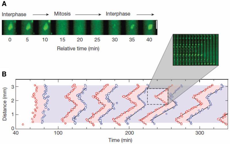
Figure 9. Waves of MPF activation in spatially extended extracts.
Data from Chang & Ferrell (2013); used by permission. (A) Cycling frog egg extracts, supplemented with sperm nuclei, are dispersed in long, thin capillary tubes. During interphase, the nuclear envelope is intact and the nuclei fluoresce green. In mitosis, when the nuclear envelope breaks down, the fluorescence disperses through the cytoplasm. As nuclei complete mitosis, the nuclear envelope re-forms and the green nuclear fluorescence re-appears. In this preparation nuclei go through repetitive mitotic cycles with a period of ~40 min. (B) Space-time plots of nuclear entry into mitosis (red points) and exit from mitosis (blue points). Over time, mitotic entry and exit organizes into traveling waves (see inset). The slope of the space-time plots is the speed of these waves, ~60 μm/min.

