Figure 5.

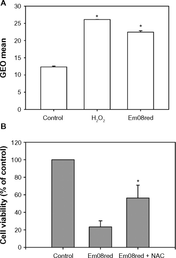
Intracellular H2O2 production accounted for em08red-mediated cytotoxicity.
Notes: (A) FaDu cells were treated with em08red (10 μM) or H2O2 (500 μM) for 24 and 16 hours, respectively, and 2′,7′-dichlorodihydrofluorescein diacetate staining was used to detect the intracellular H2O2 level. The data are presented as the geometric mean for each group and the asterisk (*) indicates a significant difference between the treated group and the control group (P<0.05). (B) The cotreatment group was preincubated with N-acetylcysteine (2 mM) for 2 hours, and em08red (10 μM) was then added to each treatment group for a further 24 hours of treatment. The control group was treated with 0.1% dimethyl sulfoxide only. The MTT assay was used to determine relative cell viability and the results are presented as the mean ± standard deviation. The asterisk (*) indicates a significant difference between the emo8red-treated group and the cotreatment group (P<0.05).
