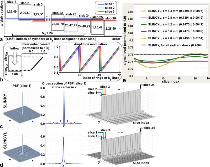
FIG. 2.
SLINKY vs. SLINCYL. a: Data reconstruction scheme of SLINCYL and SLINKY with Ns = 24 and 72 cylinders or ky lines. As highlighted with colored lines, relevant hybrid slices are selected out of Ns consecutive slabs to reconstruct each slice. b: A sawtooth shape of periodic (period = Ns) k-space amplitude modulation (right) for each colored slice in (a) caused by in flow enhancement (left). The modulation pattern is circularly shifted for different slices, which repeats every Ns slices. c,d: The magnitude of the PSF of slice 1 (left), its cross section (middle), and cross sections of the PSFs of Ns = 24 slices stacked together (right). Across the 24 slices, the PSFs of SLINCYL (d) consistently show more distributed artifacts than SLINKY (c). e: Signal levels across Ns = 24 slices with different vessel radii. Although not as constant as SLINKY, SLINCYL also provides relatively uniform signal levels for vessels with radius up to 7 mm.

