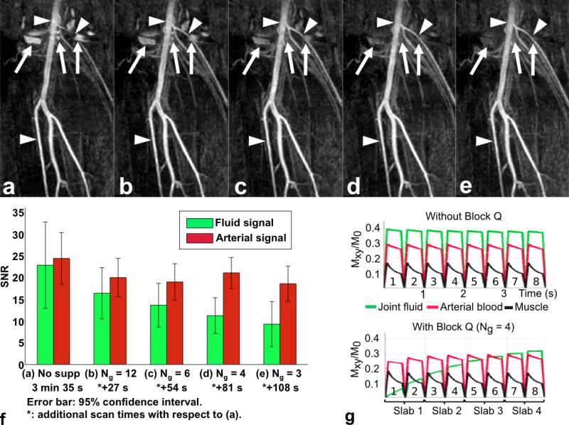
FIG. 5.
SLINCYL MRA of the calves of a healthy subject with/without block Q and corresponding Bloch simulations. Compared to the acquisition without block Q (a), the acquisitions with block Q (b: Ng = 12, c: Ng = 6, d: Ng = 4, e: Ng = 3) provide better depiction of arterial signals (arrowheads) due to the suppression of fluid signals (arrows) around the knee joint. The degree of fluid suppression is further improved as block Q is applied more frequently (i.e., with smaller Ng). f: SNR of fluid and arterial signals and scan time for each case. g: Bloch simulations of transient bSSFP signals corresponding to (a) (top) and (d) (bottom). Similar to the in vivo result, joint fluid (green) is much suppressed with block Q applied ahead of every Ng = 4 slabs, particularly for the first slab that collects the six innermost cylinders.

