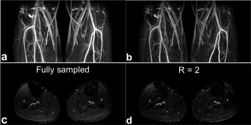
FIG. 6.
SLINCYL MRA of the calves of a healthy subject with a fully sampled and an undersampled acquisition. In addition to coronal MIP images (a,b), representative axial slice (c,d) from each acquisition is shown. The prospectively undersampled (R = 2) case reconstructed with the proposed parallel imaging method (b,d) shows comparable image quality to the fully sampled case (a,c) other than the decrease in SNR. The main arteries (popliteal, anterior tibial, posterior tibial, and peroneal) are well depicted in both cases.

