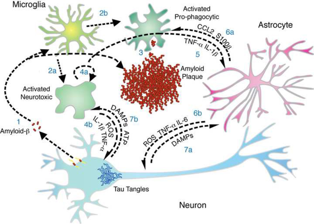
Figure 1. Inflammation in Alzheimer’s Disease.
There is a complex interplay between mediators of inflammation at sites of amyloid deposition in Alzheimer’s disease (AD). (1) Amyloid-β peptide forms aggregates and activates microglia. Microglia transition from ramified to activated, which can be (2a) neurotoxic, producing ROS, IL-1β, and TNF-α, or (2b) pro-phagocytic. (3) Phagocytic microglia will clear amyloid while neurotoxic microglia (4a) will secrete factors that act in an autocrine manner to reinforce/maintain the inflammatory phenotype and (4b) factors that directly damage neurons. (5) Factors secreted by microglia stimulate a response by astrocytes that can (6a) feedback onto microglia, driving recruitment and neuroinflammatory modulation, or (6b) act on neurons, which can be either neurotoxic or neurotrophic. Neurons, as a response to intracellular injury by tau tangles or neurotoxic ROS and cytokines, may die and release DAMPs and ATP that influence the inflammatory phenotype of (7a) astrocytes and (7b) microglia.

