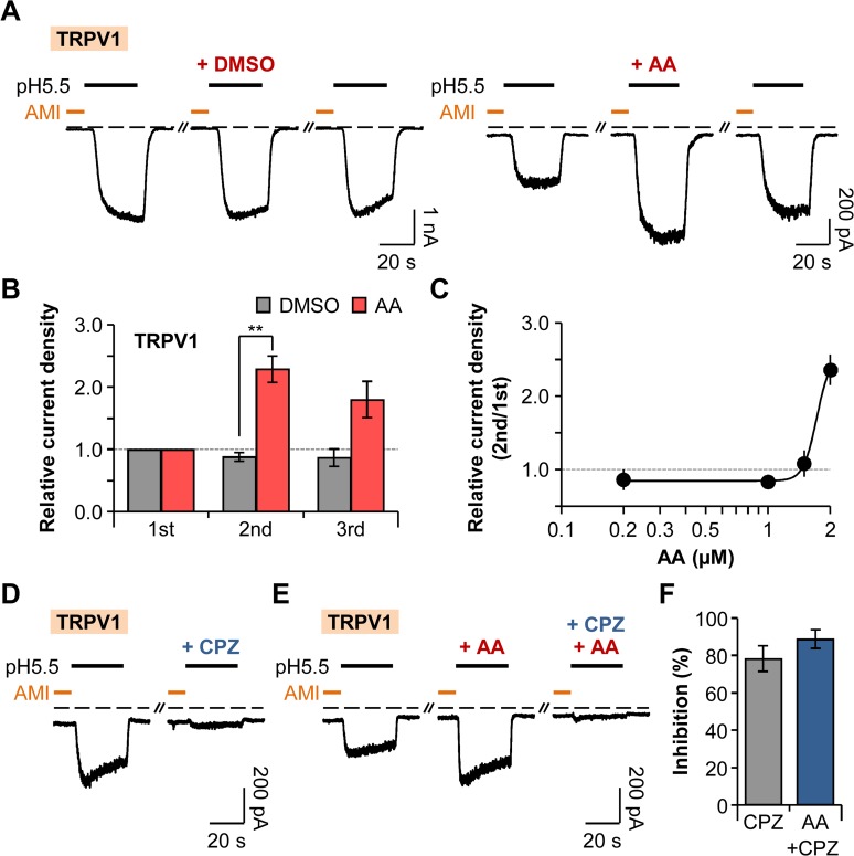
Fig 6. Potentiation of TRPV1 by AA.
(A) TRPV1 current traces repetitively activated by extracellular pH drop to 5.5 for 30 s with time intervals of 300 s. Amiloride (300 μM) was pretreated for 10 s before the pH pulses. AA (2 μM) was applied for 20 s before the second amiloride treatment. Dashed line indicates the zero current level. (B) Relative current density was measured for the cells in (A) (n = 4 for DMSO; n = 9 for AA). Current density of each pulse was divided by that of the first pulse. ** P < 0.01, with two-way ANOVA followed by Bonferroni post-hoc test and student’s t-test. (C) Dose-dependent relative current density of TRPV1 (n = 5–17). (D) TRPV1 currents were inhibited by preincubation of cells with pH 7.4 solution containing capsazepine (10 μM) for 20 s before the second amiloride treatment. (E) The potentiating effect of AA (2 μM) on TRPV1 currents was inhibited by capsazepine. (F) Percentage of inhibition by capsazepine in the absence (grey) or the presence (blue) of AA (n = 6 for CPZ; n = 6 for AA+CPZ). Data are mean ± SEM.

