Figure 8.
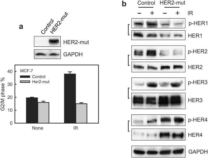
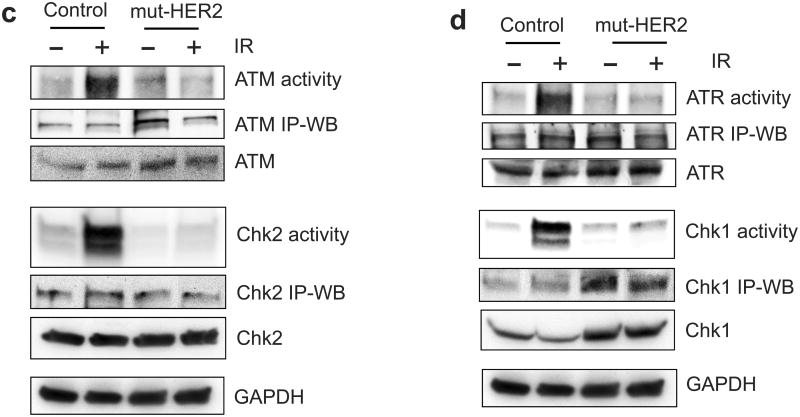
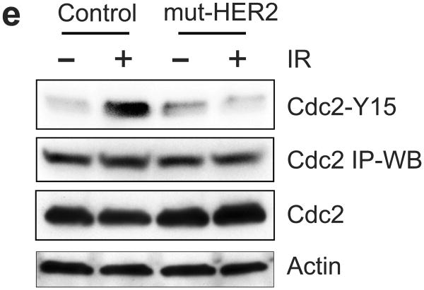
Ectopic expression of dominant negative mutant (mut) HER2 abrogates IR-induced G2/M checkpoint activation in MCF-7 cells. (a) MCF-7 cells stably transfected with a vector expressing myc-tagged HER2-mut or a relevant control empty vector were exposed to 10-Gy IR or left un-irradiated. Upper panel: The transfected cells were analyzed for HER2-mut expression by immunoblotting using anti-myc antibody. The mut-HER2 is detected as an ∼80-KD protein by Western blotting. Lower panel: the cells were exposed to 10-Gy IR, incubated for 24 h at 37°C and analyzed for cell cycle. Result depicts the percentage of cells in G2/M phase and is shown as mean±s.d. of triplicate samples, which represents two separate experiments. (b) HER2-mut expressing and control cells were exposed to 10-Gy IR and incubated for 15 min (HER1 and HER3) or 1 h (HER4). The cells were analyzed for phosphorylation and protein level of HER1, 2, 3 and 4. The levels of GAPDH in the lysates were assessed to confirm the equal protein loadings. (c) mut-HER2 expressing and control cells were exposed to 10-Gy IR, incubated for 1 h and analyzed for ATM and Chk2 activities as described above. As controls, the levels of ATM and Chk2 in the immunopreciptate and in cell lysates were analyzed by immunoblotting. GAPDH in cell lysates were probed as a protein loading control. (d) The cell samples above were analyzed for ATR and Chk1 kinase activities. As controls, the levels of ATR and Chk1 in the immunopreciptate and in cell lysates were analyzed by immunoblotting. GAPDH in cell lysates were analyzed as a protein loading control. (e) Cdc2 was immunoprecipitated from the cell samples described above in (c) and analyzed for levels of Cdc2-Y15 phosphorylation and Cdc2 protein by immunoblotting (Cdc2-Y15 and Cdc2 IP-WB). As controls, levels of Cdc2 and Actin in the cell lysates were assessed by immunoblotting.


