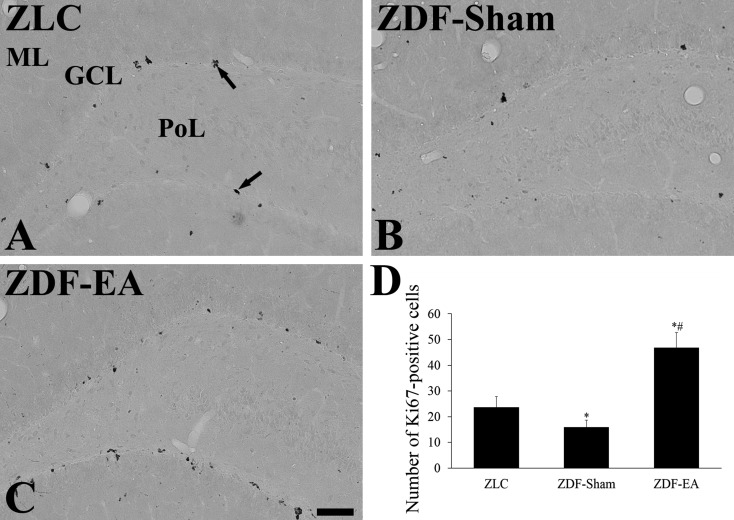
Fig. 2.
Immunohistochemistry for Ki67 in the dentate gyrus in ZLC (A), ZDF-Sham (B) and ZDF-EA (C) rats. In the ZLC group, Ki67-positive nuclei (arrows) are detected in the dentate gyrus. In the ZDF-Sham group, fewer Ki67-positive nuclei are observed in the dentate gyrus as compared to those in the ZLC group. In the ZDF-EA group, abundant Ki67-positive nuclei are detected in the dentate gyrus. GCL, granule cell layer; ML, molecular layer; PoL, polymorphic layer. Scale bar=100 µm. D: Quantitative analysis of Ki67-positive nuclei per section in the ZLC, ZDF-Sham and ZDF-EA rats using image analyzer (n=5 per group; *P<0.05, significantly different from the ZLC group, #P<0.05, significantly different from the ZDF-Sham group). Bars indicate means ± SEM.

