
Fig. 2.
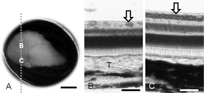
Macroscopic photograph of the ocular fundus with a normal tapetum in the right eye (A). Micrographs (B, C) taken from the parasagittal section which is cut along the dashed line. B, the thickest part of the tapetum; C, the area centralis in the visual streak. Thionine stain (B, C). T, Tapetum; Arrow, ganglion cells. Scale bar=5 mm in A and 50 µm in B and C.