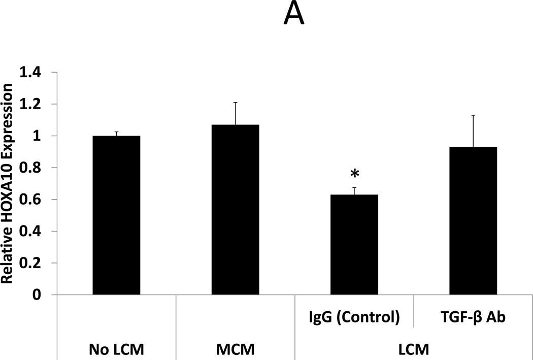
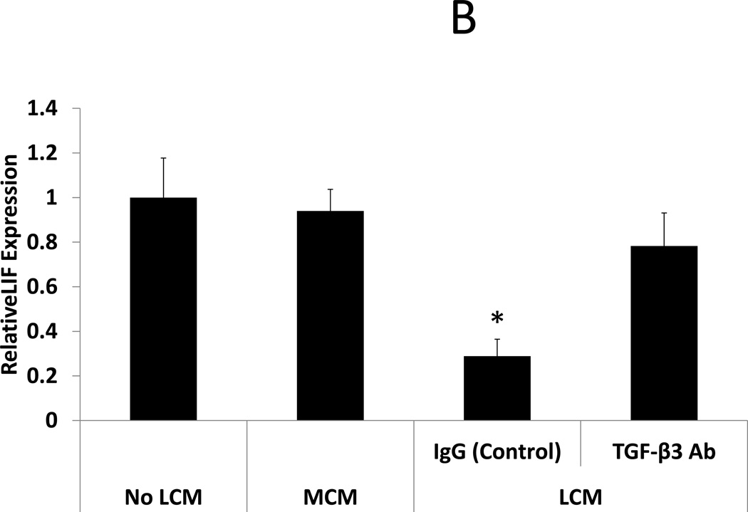
Figure 3. BMP-2 stimulated HOXA10 and LIF expression in LCM treated ESC: Effect of TGF-β Antibody.
BMP-2 responsiveness was assessed in LCM exposed ESC by treatment with rhBMP-2. HOXA10 and LIF expression were quantified by qRT-PCR after 24 hours of rhBMP-2 treatment. A: BMP-2 stimulated HOXA10 expression. BMP-2 stimulated HOXA10 expression was unchanged in MCM exposed ESC, compared to non-exposed control ESC (1.07 fold, NS). BMP-2 stimulated HOXA10 expression was repressed in ESC exposed to LCM, compared to non LCM exposed control ESC (0.63 fold, p<0.05). Incubation of LCM with TGF-β neutralizing antibody, prior to ESC exposure, prevented repression of BMP-2 stimulated HOXA10 expression. B: BMP-2 stimulated LIF expression. BMP-2 stimulated LIF expression was unchanged in MCM exposed ESC, compared to non-exposed control ESC (0.94 fold, NS). BMP-2 stimulated LIF expression was repressed in LCM exposed ESC, compared to non LCM exposed control ESC (0.29 fold, p<0.05). Incubation of LCM with TGF-β neutralizing antibody, prior to ESC exposure, prevented repression of BMP-2 stimulated LIF expression.
* denotes statistical significance with p<0.05


