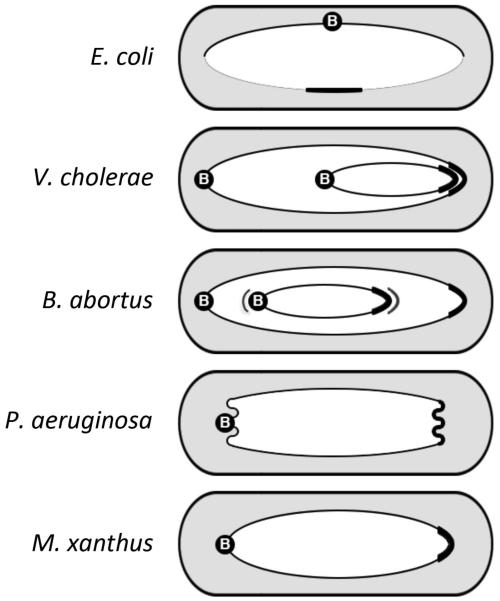
Figure 2.
Spatial arrangement of chromosomes in newborn bacterial cells. Note that the locations of origin/parS region (B) and the terminus region (capped) can vary in different bacteria. In E. coli, the terminus region is compacted by the MatP/matS system but the region also must extend to join the two arms (replichores) of the chromosome, which do not overlap under slow growth conditions. The junctions are indicated by thinner lines. In other examples, the replichores overlap over their entire length. Some other noteworthy features are as follows. The second (smaller) chromosomes in V. cholerae and in B. abortus are located differently [Deghelt et al., 2014]. The extra lines in B. abortus next to the centromere and the terminus indicate that their locations are not that fixed and move around their mean positions a bit. In P. aeruginosa, the origin and terminus regions are more compacted than in other bacteria [Vallet-Gely and Boccard F, 2013]. Both regions are also more removed from the poles. In M. xanthus, the regions are also removed from the pole. It is worth noting that centromeres are poleward in all examples (an exception is E. coli which may not have a centromere).

