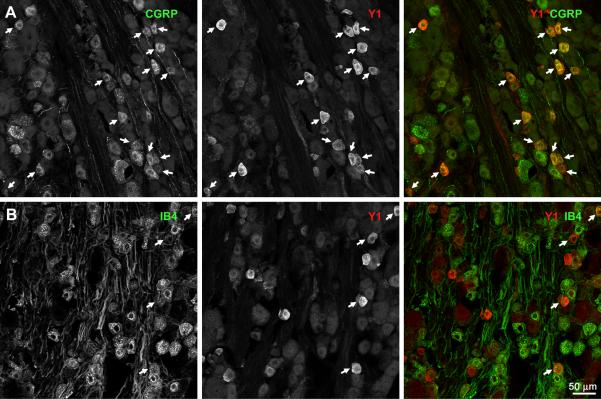
Fig. 3. Presence of Y1 receptors in rat DRG neurons.
Sections from lumbar DRG (L1–L6) were labeled with antibodies against CGRP or the Y1 receptor, or with isolectin B4-biotin (IB4). Each image consists of 4 confocal sections spaced 0.99 μm, taken with a 20x objective. A) CGRP and Y1 receptor immunoreactivities. B) IB4 staining and Y1 receptor immunoreactivity. Arrows indicate double-labeled cell bodies.

