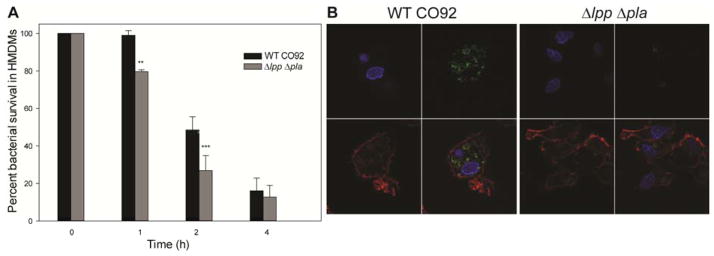
Figure 4. Intracellular survival of WT Y. pestis CO92 and its mutant strains in human monocyte-derived macrophages (HMDMs).

Macrophages from three individual donors were infected at an MOI of 0.5 with either the WT CO92 or Δlpp Δpla double mutant for 40 min following 1 h gentamicin treatment (10 μg/ml). At 0, 1, 2, and 4 h (post-gentamicin treatment), macrophages were harvested and the number of bacteria inside macrophages was counted. The percent survival rate was calculated based on the number of bacteria recovered at each specific time point over that of the 0 h time point. The pooled data were shown and error bars indicated standard deviations for percent intracellular bacterial survival (A). The data were analyzed using One-way ANOVA with Bonferroni correction and p values ≤0.05 were considered significant. Asterisks indicate statistically significant differences compared to WT CO92-infected HMDMs (**p<0.01, ***p<0.001). At the 1 h time point, the infected HMDMs were also fixed and the bacteria inside the macrophages were detected by specific anti-F1 (capsular antigen) antibodies follow by secondary Alexa Fluor 488 antibodies (green). The actin filaments were stained with Alexa Fluor 568 phalloidin (red), and the cell nucleus was visualized with DAPI (blue). The images were taken by confocal microscopy with the magnification of 1,000 (B).
