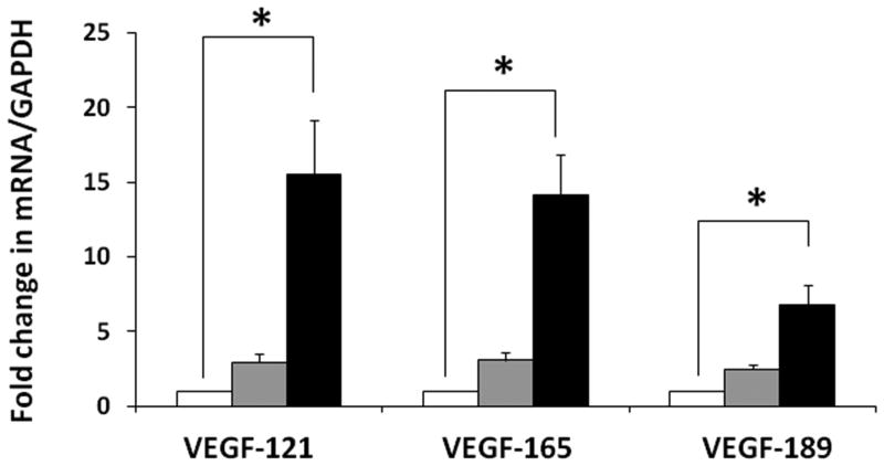
Figure 6. Developmental change in VEGF-A splice variants in the midgestation fetal retina.
Bar diagrams (means ± SE) show fold change in the mRNA expression of VEGF-A splice variants, VEGF121, VEGF165, and VEGF189, in retinal tissue from fetuses of 10-14 weeks, 15-19 weeks, and 20-24 weeks. * indicates P<0.05.

