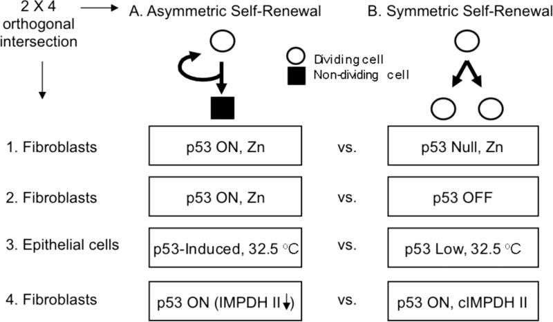
Figure 1. A 2 × 4 orthogonal-intersection microarray analysis to detect genes associated with asymmetric self-renewal.
cDNA micro-arrays were developed as described in Materials and Methods for four distinct comparisons of cells in congruent states of asymmetric self-renewal (A) versus symmetric self-renewal (B). In this strategy, asymmetric self-renewal associated (ASRA) genes [5] are defined as those whose expression consistently changes by a significant degree between asymmetric self-renewal versus symmetric self-renewal states in all four self-renewal pattern comparison models. Comparison 1 paired asymmetrically self-renewing, p53-induced Ind-8 MEFs to symmetrically self-renewing, p53-null Con-3 MEFs, both with ZnCl2-supplemention (Rambhatla et al., 2001; Rambhatla et al., 2005). Comparison 2 paired asymmetrically self-renewing, p53-induced Ind-8 cells (ZnCl2-supplemented) to symmetrically self-renewing, non-induced Ind-8 cells (ZnCl2-free). Comparison 3 paired asymmetrically self-renewing, p53-induced 1h-3 mouse mammary epithelial cells to symmetrically self-renewing, p53-expressing 1g-1 cells, both grown at 32.5°C (Sherley et al., 1995a; Rambhatla et al, 2001; Rambhatla et al, 2005). Culture of 1h-3 cells at 32.5°C causes a 1.5-fold elevation in p53 protein expression and a shift to asymmetric self-renewal due to a stably transfected low temperature-inducible p53 mini-gene. Line 1g-1 cells are congenic control cells that have subnormal p53 expression, lack the inducible p53 cDNA sequences, and maintain symmetric self-renewal at 32.5°C. Comparison 4-paired two stably transfected derivatives of Ind-8 cells under conditions of ZnCl2-supplementation. Line tC-2, which was derived by transfection with only gene expression regulatory elements, retains asymmetric self-renewal like the parental Ind-8 cells. In contrast, tI-3 cells, which were derived with synonymous expression constructs that contained a hamster type II inosine-5’-monophosphate dehydrogenase (IMPDH II) cDNA, no longer shift to asymmetric self-renewal with ZnCl2-supplemention (Liu et al., 1998a; Rambhatla et al., 2005). In tC-2 cells, IMPDH II protein levels are reduced as a result of p53 expression. In tI-3 cells, although the endogenous IMPDH II is reduced by p53 expression as well, the overall cellular level of IMPDH II is maintained by the constitutive expression the IMPDH II transgene (cIMPDH II) (Liu et al., 1998a).

