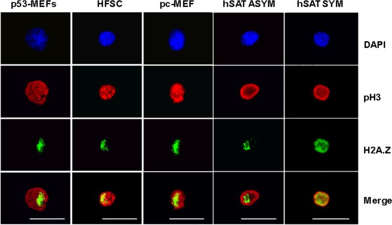
Figure 2. H2A.Z asymmetry is detected in prophase nuclei, identified by phosphorylated histone H3, for a variety of cultured cell types.
p53-MEFs, example of H2A.Z asymmetry in genetically engineered mouse embryo fibroblasts (line Ind-8) (Rambhatla et al., 2001; Rambhatla et al., 2005; Liu et al., 1998b) under conditions that promote asymmetric self-renewal. HFSC, example of H2A.Z asymmetry in mouse hair follicle stem cells (strain 3C5) (Huh et al., 2011; Huh and Sherley, 2011) under conditions that promote asymmetric self-renewal. pc-MEF, example of cell with H2A.Z asymmetry detected in cultures of pre-crises MEFs (See also Fig. 3). In cultures enriched for human skeletal muscle satellite stem cells: hSAT ASYM, example of cell with H2A.Z asymmetry, and hSAT SYM, example of cell with symmetric H2A.Z. DAPI, nuclear DNA fluorescence. pH3, indirect in situ immunofluorescence (ISIF) with antibodies specific for phosphorylated histone H3. H2A.Z, ISIF with antibodies specific for H2A.Z. ISIF was performed simultaneously for pH3 and H2A.Z. Merge, overlaid anti-pH3 and anti-H2A.Z images. Scale bar = 25 microns.

