
Figure 1.
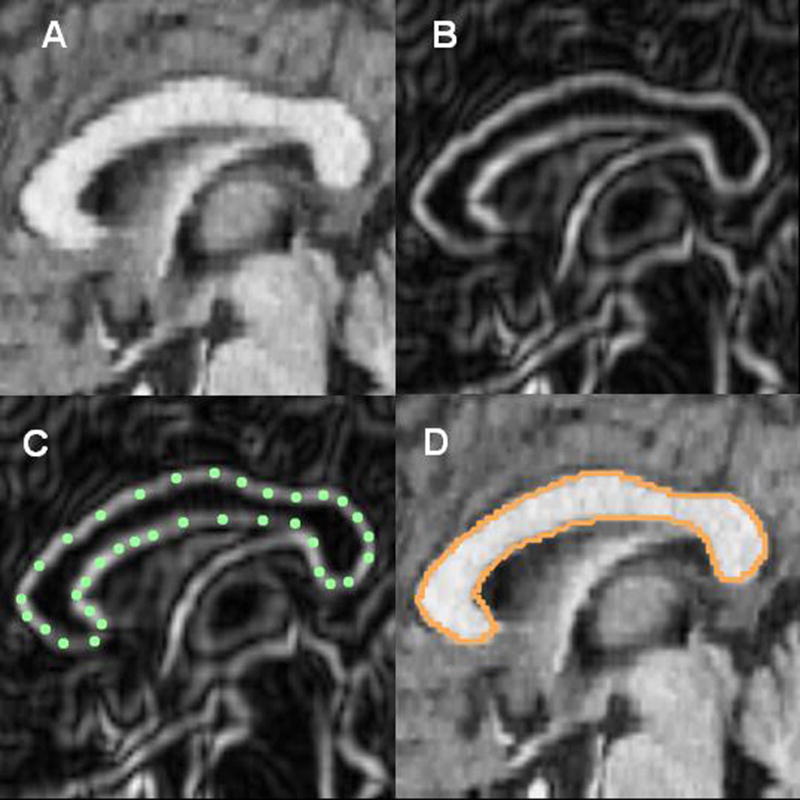
Tracing the corpus callosum. (A) Magnetic resonance image showing a sagittal cross-section through the corpus callosum. (B) Using an edge contrast-enhancing technique (Sobel-gradient filter), gray/white boundaries of the corpus callosum are enhanced. (C) the corpus callosum is outlined; and (D) the spline curve is fit.