Figure 5.
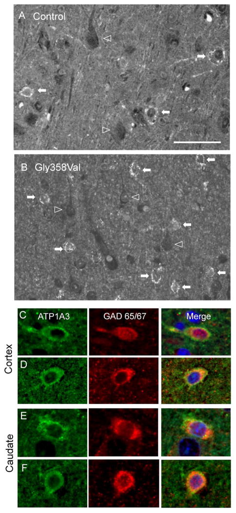
ATP1A3 immunofluorescence was localized to GABAergic cells in human temporal cortex and basal ganglia. A,B) Control (A) and ATP1A3 Gly358Val (B) cortex. Filled arrows indicate cells with interneuron morphology and perisomatic ATP1A3 IF; open arrowheads indicate outlines of cells with pyramidal morphology and absence of ATP1A3 IF. C,D) ATP1A3 (green) co-localizes with the interneuron marker GAD 65/67 (red) in control (C) and Gly358Val (D) human cortex. E,F) ATP1A3 colocalization with GAD 65/67 in human caudate nucleus. Scale bars: 50 μM

