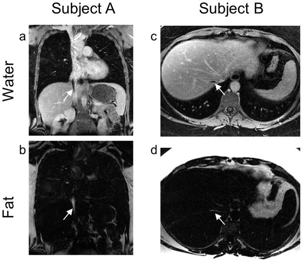
FIG 4.
Flow-induced fat-water signal misallocations may occur in vivo when spins flow along the readout direction. Two such instances are shown here. In Subject A, the readout direction is Superior/Inferior in this coronal acquisition, and in Subject B, the readout direction is Right/Left in this axial acquisition. Note the dark spot (arrow) in the water image from Subject A resembling an IVC clot and the dark spot (arrow) in the water image from Subject B resembling a hepatic vein thrombus similar to that found in patients with Budd-Chiari syndrome. The bright signal missing from the water images is clearly seen in the fat images (arrows). A Gadolinium-based Contrast Agent (GBCA) was administered prior to acquisition of these fat-water-separated images.

