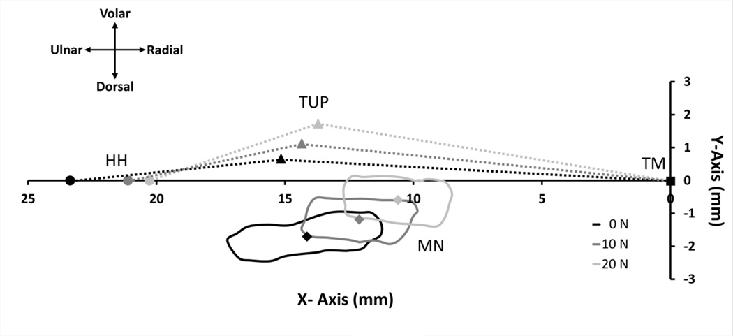
Figure 3.
The displacement of the hook of hamate (HH,  ), thenar muscles ulnar point (TUP,
  ), and median nerve (MN, solid line) with centroid (
 ) for a representative subject at 0, 10, and 20 N of wrist compression relative to the anatomically defined coordinate system with its origin at the trapezium (TM,
  ). The area beneath the dotted lines bounded by the X-Axis represents the carpal arch area.

