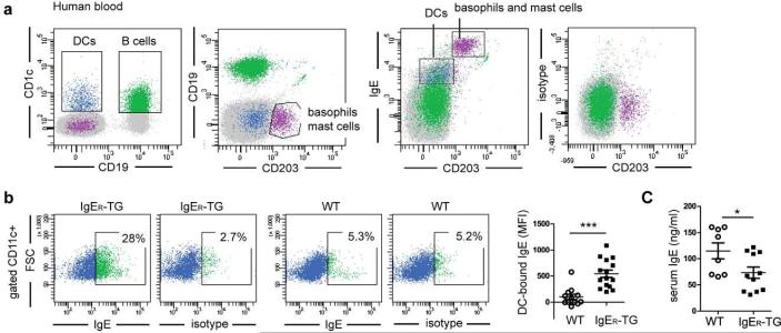
Figure 1.
Expression of FcεRI on DCs results in a DC-specific IgE pool as seen in non-allergic humans. (a) Human peripheral blood DCs of a non-allergic individual carry surface IgE. DCs (in blue) were identified by gating on CD1c+ CD19− cells to distinguish them from CD1c+CD19+ B cells (in green) and analyzed for cell surface-bound IgE. DCs carry significant amounts of IgE but less than CD203+ basophils and mast cells (in purple). (b) CD11c+ DCs of IgER-TG mice carry IgE at steady state. Representative FACS plots of WT- and IgER-TG DCs isolated from spleens and quantification of the DC-bound IgE pool using mean fluorescence intensities (MFI) determined by flow cytometric analysis. (c) Baseline serum IgE levels are lower in non-sensitized IgER-TG animals. Symbols in are representative of individual mice of ≥ 2 independent experiments (*p < 0.05; ***p< 0.001).

