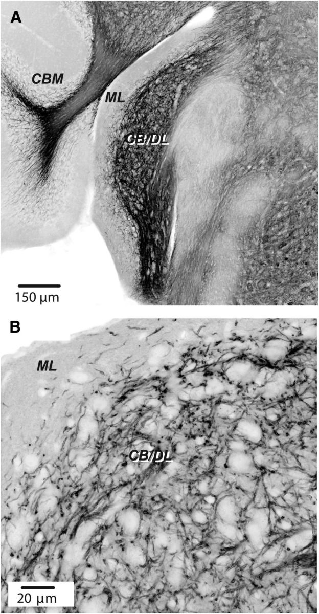
Figure 7.

Expression of ChR2 in DCN in a Tph2-ChR2-EYFP mouse line. A, Section showing DCN and adjoining structures. Image printed in negative shows fluorescent labeling using antibody labeling for EYFP. ML, Molecular layer of DCN; CB/DL, fusiform cell body and deep layers; CBM, cerebellar cortex. Confocal image taken with a 10× objective. B, Labeling in DCN imaged using a 60× oil-immersion objective. Confocal image stack of four planes. Note preponderance of labeling up to the cell body layer and relative absence in the molecular layer.
