FIG 2.
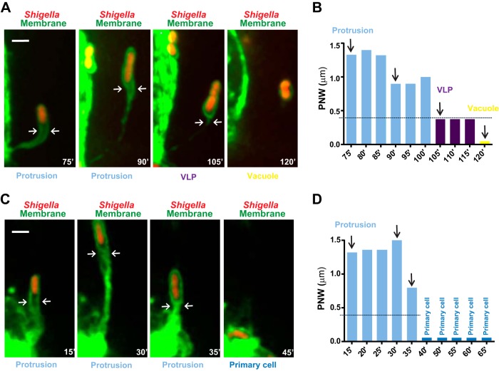
S. flexneri spreads from cell to cell in HT-29 cells through vacuole-like protrusion (VLP) formation. Time-lapse microscopy of HT-29 cells (green, GFP membrane marker) infected with RFP-expressing S. flexneri (red, Shigella). (A) Representative images showing a bacterium forming a membrane protrusion (Protrusion) that transitions into a vacuole-like protrusion (VLP), which further resolves into a vacuole (Vacuole). Bar, 2 μm. (B) Graph showing the quantification of the protrusion neck width (PNW; arrows in panel A) in images from Movie S1 in the supplemental material (75′ to 120′) and corresponding images (vertical arrows) as shown in panel A. Protrusion, light blue, PNW of >0.4 μm; VLPs, purple, PNW of <0.4 μm; vacuole yellow, no PN. (C) Representative images showing a bacterium forming a membrane protrusion (Protrusion) that fails to transition into a vacuole-like protrusion and retracts to the primary infected cell (Primary cell). Bar, 2 μm. (D) Graph showing the quantification of the protrusion neck width (PNW; arrows in panel A) in images from Movie S2 in the supplemental material (15′ to 65′) and corresponding images (vertical arrows) as shown in panel C. Protrusion, light blue, PNW of >0.4 μm; Primary cell, dark blue.

