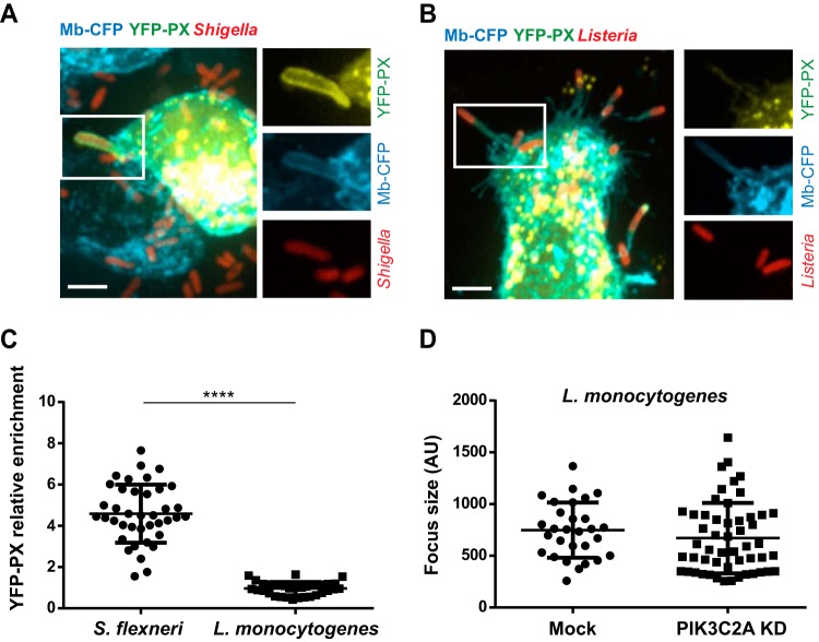
FIG 6.
L. monocytogenes dissemination does not rely on PtdIns(3)P production in HT-29 cells. (A and B) Representative images of protrusions formed in CFP membrane marker-expressing HT-29 cells expressing the YFP-tagged PtdIns(3)P-binding PX domain construct and infected with RFP-expressing S. flexneri (A) or L. monocytogenes (B) for 4 h. Bar, 5 μm. (C) Graph showing statistical analyses of the relative enrichment of the YFP-PX probe in protrusions formed by S. flexneri or L. monocytogenes in HT-29 cells (****, P < 0.0001; unpaired t test). (D) Graph showing the quantification of L. monocytogenes infection focus size in mock-treated and PIK3C2A-depleted HT-29 cells 8 h postinfection.

