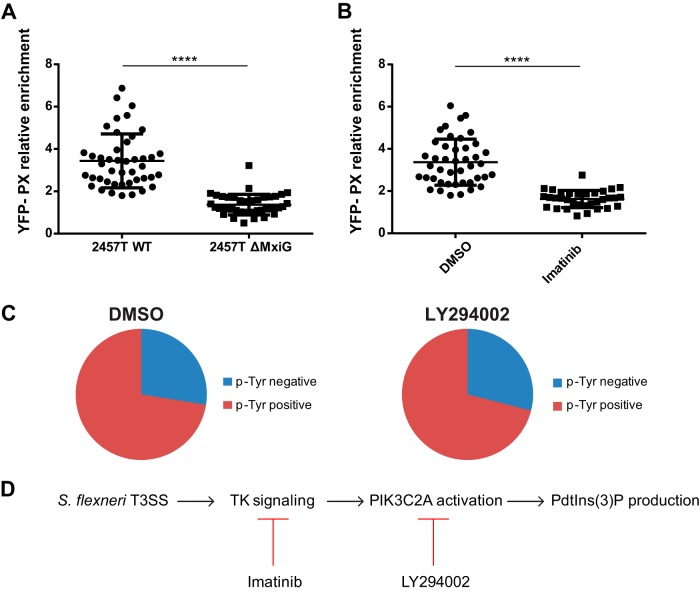
FIG 7.
PtdIns(3)P production relies on the bacterial type 3 secretion system and host cell tyrosine kinase signaling in HT-29 cells. (A) Graph showing statistical analyses of the relative enrichment of the YFP-PX probe in HT-29 cells infected with wild-type S. flexneri (2457T WT) or ΔMxiG mutant (2457T ΔMxiG) (****, P < 0.0001; unpaired t test). (B) Graph showing statistical analyses of the relative enrichment of the YFP-PX probe in DMSO-treated (DMSO) and HT-29 cells treated with imatinib (100 nM) (Imatinib) and infected with wild-type S. flexneri (****, P < 0.0001; unpaired t test). (C) Graph showing the quantification of phosphotyrosine-positive and -negative protrusions in DMSO- and LY294002-treated HT-29 cells (10 μM) infected with wild-type S. flexneri (DMSO versus LY294002, P > 0.1; unpaired t test). (D) VLP formation in HT-29 cells is mediated by a signaling cascade that relies on the T3SS and activation of tyrosine kinase signaling in protrusions and the subsequent PIK3C2A-dependent production of PtdIns(3)P.

