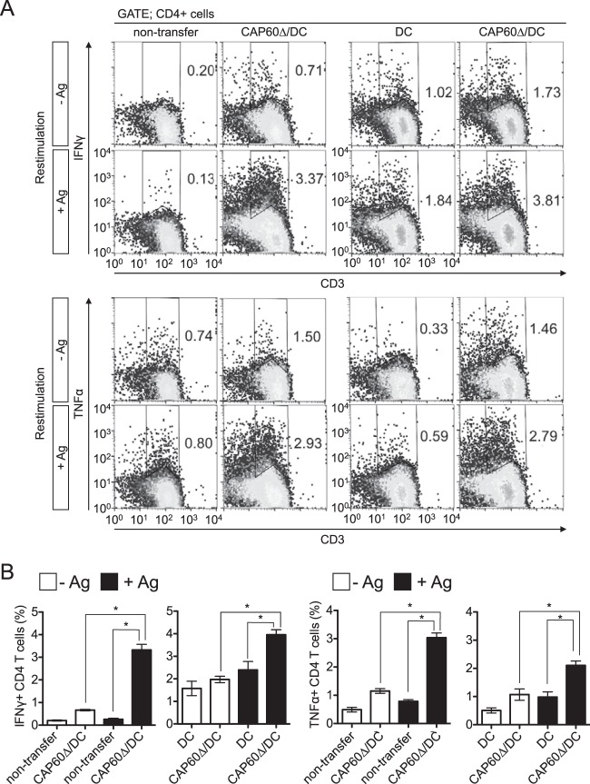
FIG 4.
Transferring CAP60Δ/DCs induces cytokine-producing CD4 T cells in spleen. Splenocytes were obtained from three mice at 14 days after infection and cultured with (+Ag) or without (−Ag) antigen and with or without heat-killed Δcap60 cells (MOI = 0.1) for 5 to 6 days. For flow cytometry analysis, gates were set for CD4+ cells. Representative flow cytometry profiles (A) and histograms for statistical analysis (B) are shown. Pooled data from two separate experiments were used to prepare histograms (means ± SEMs). *, P < 0.05 by analysis of variance with Dunnett's post hoc test.

