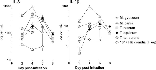
FIG 2.
Dermatophyte infection results in cytokine release by EpiDerm tissues. Tissues were infected with 106 conidia from the five dermatophyte species, as described for Fig. 1. The control is 106 heat-killed conidia of T. equinum. Time points were chosen for each species that encompassed the peak LDH release determined in Fig. 1. Cytokines IL-8 and IL-1β were detected using the cytometric bead array kit. Data points represent the averages from three biological triplicates ± standard errors of the mean (SEM). For IL-8, the results with all live species conidia were statistically different (P < 0.05) from heat-killed conidia at days 2 and 4 (and all species except M. canis at 6 days). The results for M. gypseum conidia were significantly higher (P < 0.05) than those for all other species on days 2 and 4. For IL-1β, the results with all live species conidia were statistically different (P < 0.05) from heat-killed conidia on days 2 and 4 (and all species but M. canis at 6 days). The results for M. gypseum conidia were significantly higher (P < 0.05) than those for all other species on days 2 and 4.

