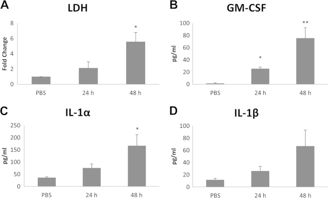
FIG 3.
Dermatophyte infection of primary keratinocytes causes damage and cytokine release. T. equinum infections were performed for three separate donor isolations of keratinocytes with 106 conidia for 24 and 48 h. (A) Detection of LDH in spent medium; (B to D) detection of cytokines by Luminex microbead assay in spent medium at 24 and 48 h postinfection. Data points represent the averages from three biological triplicates with separate donor primary keratinocytes ± SEM. Statistics were performed comparing postinfection cells with uninfected, 0-h-time-point cells. *, P < 0.05; **, P < 0.01.

