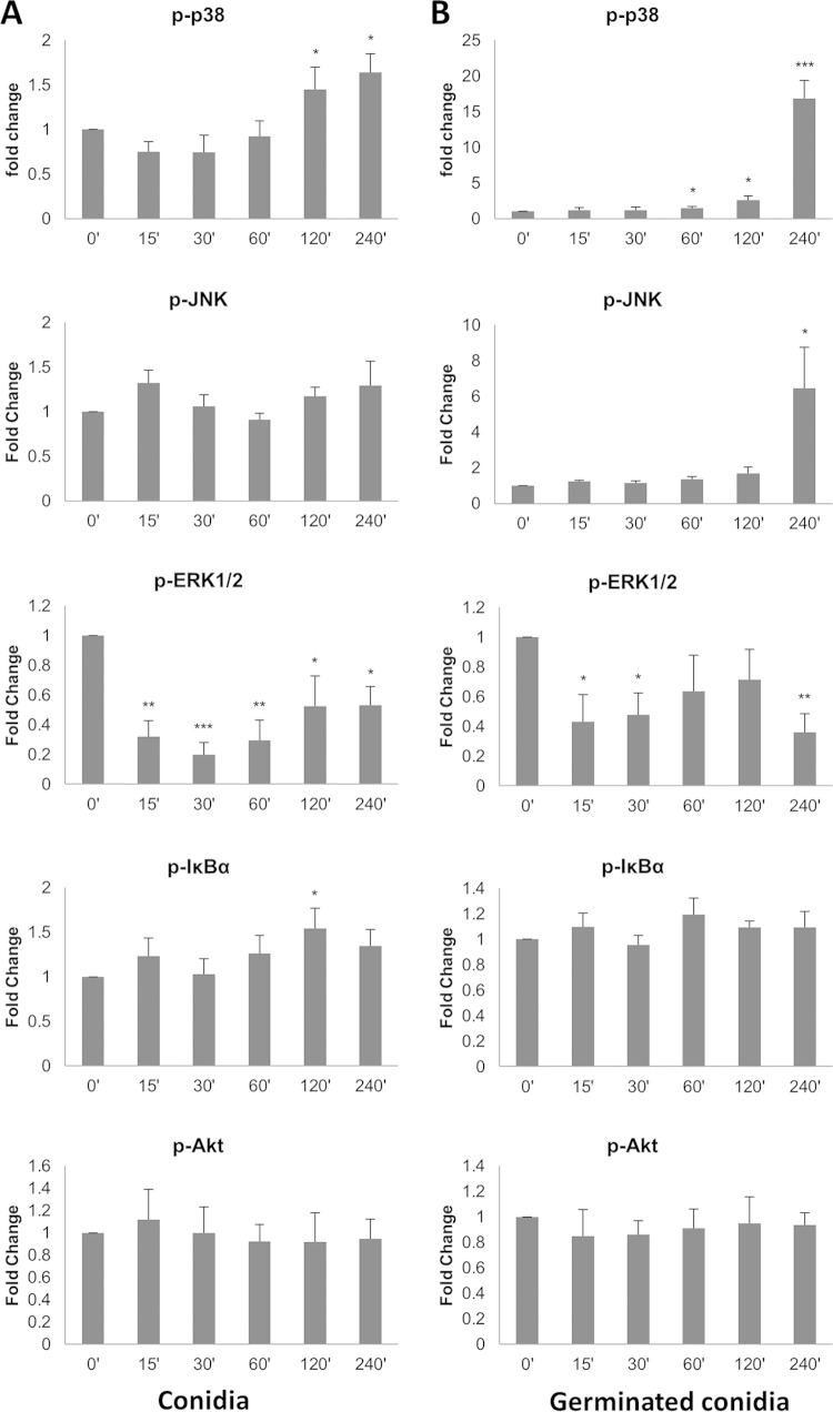
FIG 4.
Dermatophyte infection of primary keratinocytes activates MAPK signal pathways. T. equinum infections were performed for three separate donor isolations of keratinocytes with 106 conidia (A) or pregerminated conidia (B). Data points represent the averages from three biological triplicates with separate donor primary keratinocytes ± SEM. Statistics were performed comparing postinfection cells with uninfected, 0-h-time-point cells. *, P < 0.05; **, P < 0.01; ***, P < 0.001.

