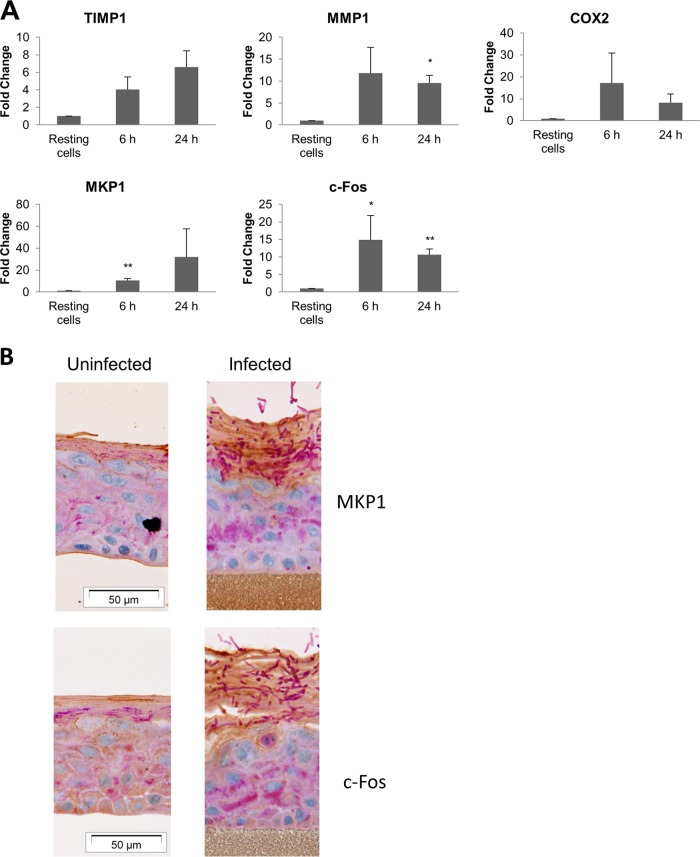
FIG 5.
Dermatophyte infection of EpiDerm tissues results in expression of tissue remodeling and proinflammatory genes and MAPK signaling proteins. T. equinum infections were performed in triplicate with germinated or ungerminated conidia. (A) Expression of tissue remodeling (timp1 and mmp1), proinflammatory (cox2), signal pathway regulatory (mkp1), and transcription factor (cfos) genes after infection. Data points represent the averages from three biological triplicates ± SEM. Statistics were performed comparing postinfection cells with uninfected, 0-h-time-point cells. *, P < 0.05; **, P < 0.01. (B) Expression of the MAPK pathway-associated regulator, MKP1, and the MAPK transcription factor, c-Fos, in infected EpiDerm tissue 96 h postinfection.

